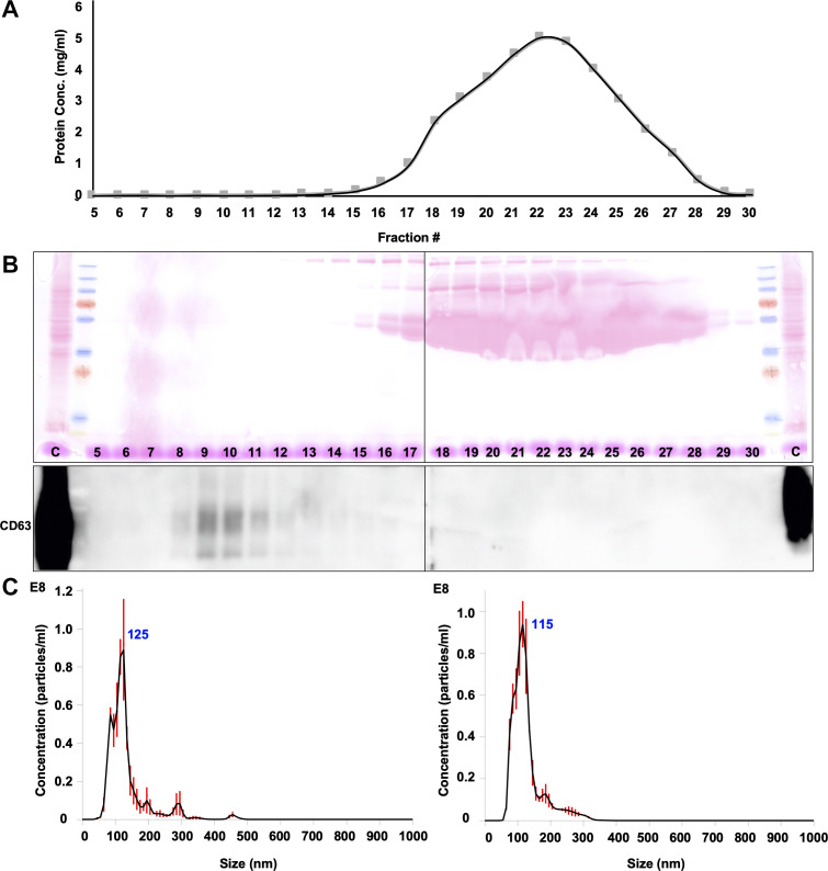
Fig. 1.
Exosome isolation using size exclusion chromatography (SEC). Endothelial cells were cultured in medium containing 5% exosome-depleted FBS. The supernatant was collected and ultrafiltered after 24 h and then processed in SEC with 10 mL of bed volume. Each 500 μL of fraction was collected after the sample was added on top of the column. A: protein concentration of each fraction was determined using Nanodrop. B: Western blot was performed using 2 separate gels to detect the distribution of vast number of proteins and exosomes in fraction 5–30. Ponceau red stain confirmed that the vast number of proteins were eluted in later fractions, whereas anti-CD63 detected exosomes in early fractions. C, parent cell lysates as positive control. C: nanoparticle tracking analysis confirmed the exosome sizes in F9 (left) and F10 (right).

