Fig. 4.

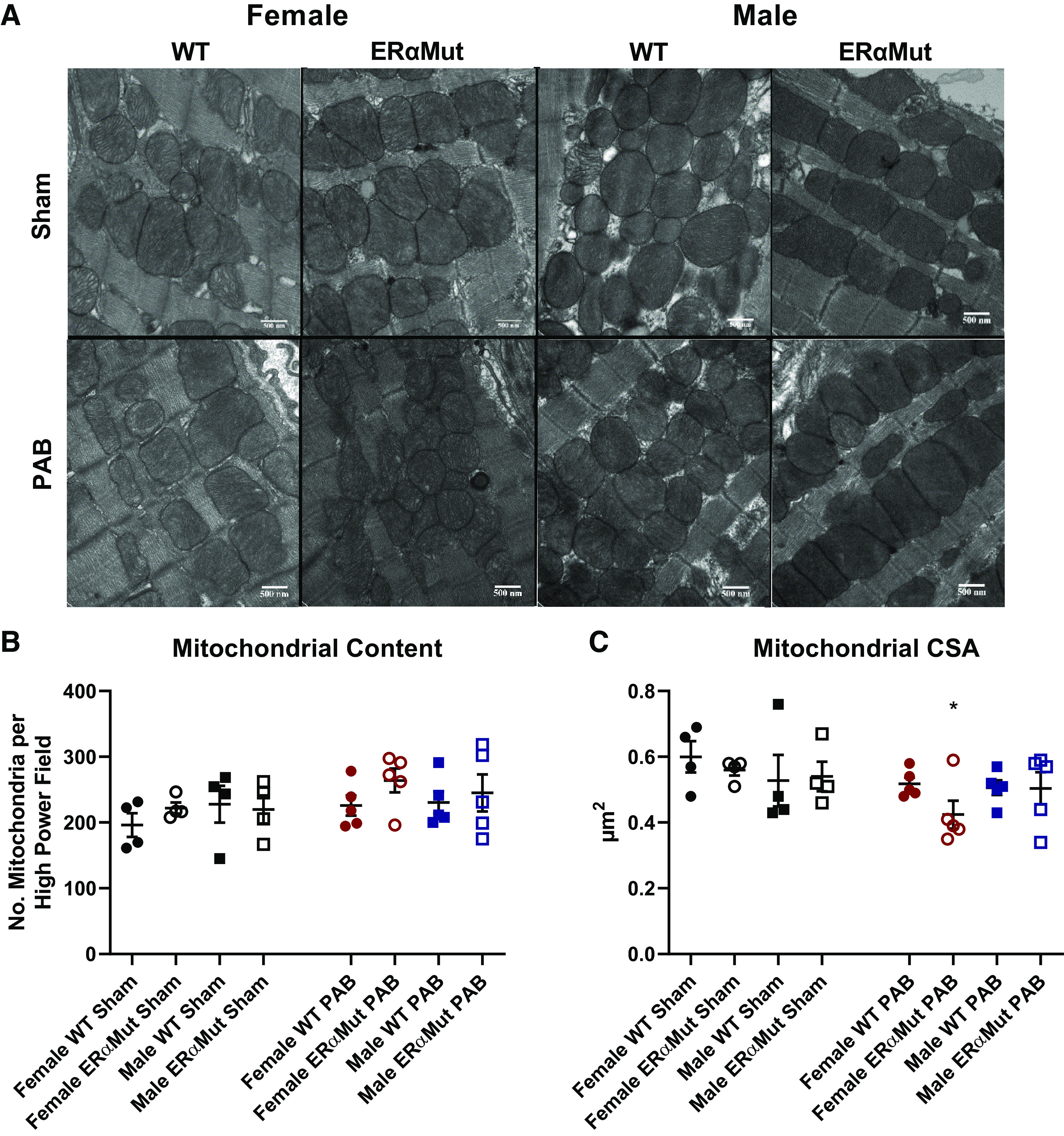
Loss of functional ERα reduces RV mitochondrial size in females with PAB. Effects of sex, ERα mutation, and PAB on mitochondrial content (the number of mitochondria) (B) and mitochondrial cross-sectional area (CSA) (C) are shown. Note the lack of change in mitochondrial content across all groups in response to PAB and the reduced mitochondrial size only in the female ERαMut rats. A: representative electron micrographs are shown of the mitochondria in the RV tissue at ×25,000 magnification. Data are presented as means ± SE. *P < 0.05 PAB vs. Sham of same sex and genotype (two-way ANOVA with Bonferroni test for multiple comparison). n = 4−5 for each group. ERα, estrogen receptor α; ERαMut, loss-of-function mutation of ERα; PAB, pulmonary artery banding; RV, right ventricular.
