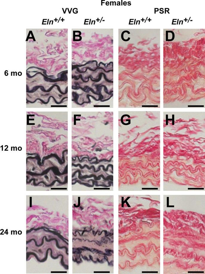
Fig. 6.

Representative female right common carotid (RCC) histology images stained with Verhoeff Van Gieson (VVG); A, B, E, F, I, and J) or picrosirius red (PSR; C, D, G, H, K, and L) for Eln+/+ (A, E, I, C, G, and K) and Eln+/− (B, F, J, D, H, and L) mice at 6 (A–D), 12 (E–H), and 24 (I–L) months of age. Images from male mice are similar; 5–7 images were examined per group. Scale bar = 20 µm.
