
Fig. 1.
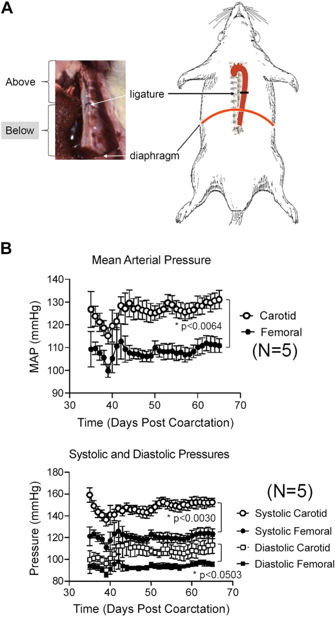
A: photograph and diagram of ligature placement on the descending thoracic aorta of the rat. B: radiotelemetric measures of mean (top) and systolic/diastolic (bottom) pressures in coarcted male Sprague–Dawley rats for 1 mo after implantation of a dual radiotelemeter (carotid for pressure above the ligature, femoral for pressure below). Points are means + SE for N number of individual rats. *Significant differences between bracketed groups as determined by mixed-effect analysis.