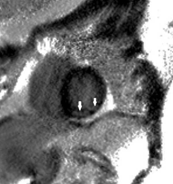
Figure 7. – Short- axis delayed contrast-enhanced PSIR cardiac MR image, showing focal subendocardial transmural and subepicardial enhancement areas, mostly in the inferior and inferolateral left ventricular walls (arrows), indicating scar tissues in the distribution of the LAD.

