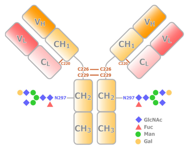
Figure 1.
Schematic representation of the structure of IgG. An IgG consists of two heavy chains (yellow) and two light chains (red). The VH and CH1 domains from a heavy chain interact with a light chain to form a Fab portion, while CH2 and CH3 domain dimer forms Fc portion. Fab and Fc portions are connected by the hinge region, where multiple conserved disulfide bridges are present. CH2 domain has a conserved N-glycosylation site.

