Figure 2.
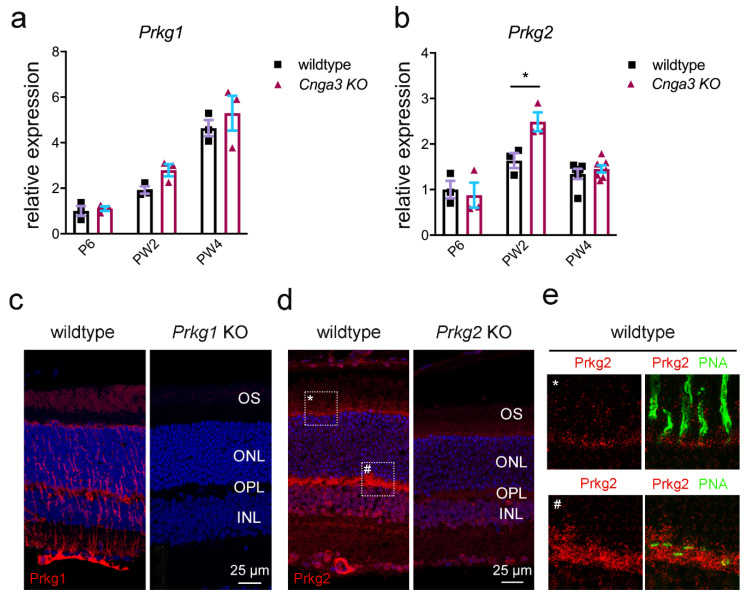
Expression of the cGMP dependent kinases Prkg1 and Prkg2 in wildtype and Cnga3 KO retina. (a,b) Relative Prkg1 (a) and Prkg2 (b) transcript levels at indicated time points. While Prkg1 is expressed at similar levels in wildtype and Cnga3 KO retina (a), there is a transient significant upregulation of Prkg2 transcript in the Cnga3 KO retina around eye opening (PW2) (b, n = 3–6, * p < 0.01, 1-way-ANOVA). (c–e) Immunolocalization signal for Prkg1 (c) and Prkg2 protein (d,e) in the mouse retina. (c) The intense Prkg1 signal (red) is mostly confined to Müller glia cells and blood vessels with additional faint signal in photoreceptor outer segments (OS). (d) Intense Prkg2 signal is found in photoreceptor inner segments (see higher magnification view on marked regions in panel (e)), synapses in the outer plexiform layer (OPL), some inner nuclear layer (INL) cells and in blood vessels. Panels c and d show Hoechst nuclear dye signal in blue and panel E shows the cone marker peanut agglutinin (PNA) signal in green. Tissue from the corresponding knockout mouse lines (Prkg1 KO in C and Prkg2 KO in (d)) did not produce a signal similar to the wildtype. The scale bar in c and d applies to both images, respectively. (e) Magnification view of the corresponding regions (*,#) marked with dotted rectangles in the left (wildtype) image of (d).Error bars shown are SEM. ONL, outer nuclear layer; P, postnatal day; PW, postnatal week.

