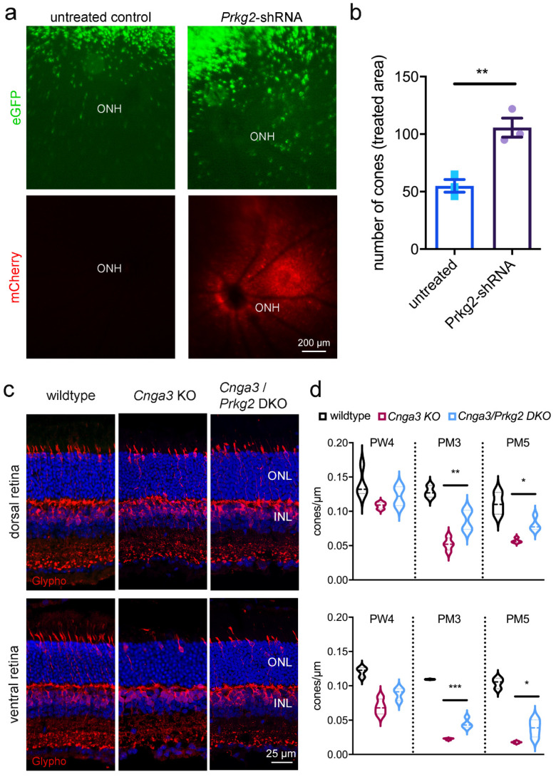
Figure 3.
Downregulation or knockout of Prkg2 delays cone degeneration in Cnga3 KO mice. (a,b) Subretinal administration of AAV vectors encoding a shRNA directed against Prkg2 preserves the number of cones (green) in RG-eGFP/Cnga3 KO mice. The retinal area targeted by the subretinal injection of the AAV vector is indicated by the fundus fluorescence signal of the vector-encoded mCherry (red) (lower panel in a). The scale bar applies to all images. (b) Quantification of the number of eGFP-positive cones from fundus fluorescence images revealed a significant preservation upon AAV-Prkg2-shRNA treatment at 3 months after injection (n = 3, ** p < 0.01 Student’s t-test). Error bars shown are SEM. ONH, optic nerve head. (c,d) Genetic inactivation of Prkg2 preserves Cnga3-deficient cone photoreceptors. (c) Cone morphology in wildtype, Cnga3 KO and Cnga3/Prkg2 double knockout (DKO) retina visualized by immunolabeling of glycogen phosphorylase (glypho). The anti-glypho signal is found in cone inner segment, cell body and synapse. In addition, anti-glypho labels bipolar cell bodies and synapses in the outer nuclear layer (ONL) and inner plexiform layer. Panels in C show Hoechst nuclear dye signal in blue. The scale bar applies to all images. (d) Violin plot showing quantification of cone numbers from glypho labelled retinal cross-sections revealing a significantly higher density of cones in Cnga3/Prkg2 DKO compared to Cnga3 KO (n = 3–4, * p < 0.05, ** p < 0.01, *** p < 0.005, 1-way-ANOVA). INL, inner nuclear layer; PM, postnatal month; PW, postnatal week.

