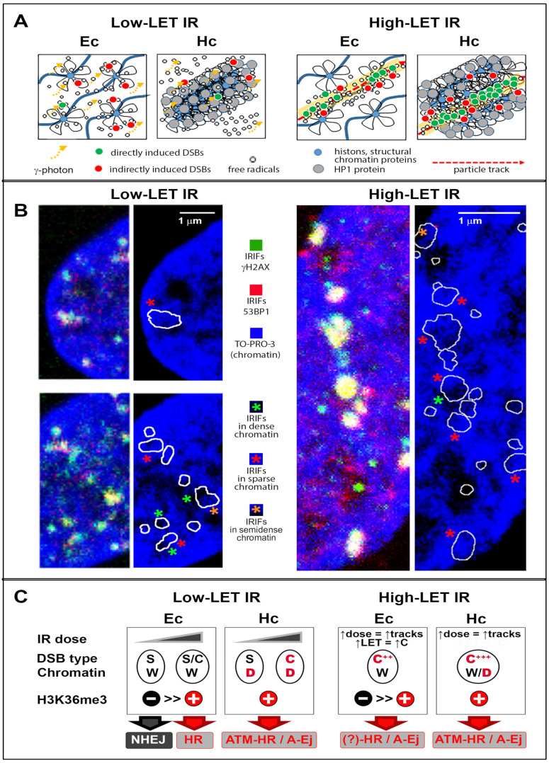
Figure 3.
The relationship between the physical properties of the incident radiation, chromatin architecture, character of DSB damage and repair mechanism. (A) Euchromatin (Ec) and heterochromatin (Hc) interact differently with ionizing radiation. This interaction is also dependent on the physical parameters of the incident radiation, especially its linear energy transfer (LET). After low-LET irradiation, highly compacted heterochromatin is better protected than euchromatin against indirect radiation DNA damage due to its lower hydration, higher occupation by chromatin-binding proteins, and, in turn, lower accessibility of DNA to harmful free radicals (e.g., [76]). However, heterochromatin is the more critical target for high-LET particles that (mostly) damage DNA directly. In contrast to the indirect damage mediated by radicals, the damage generated by concentrated energy release from high-LET particles cannot be prevented by the condensed, protein-rich heterochromatin architecture. As heterochromatin offers more DNA targets per unit than euchromatin, its interaction with energetic particles leads to the formation of more complex DSBs than in euchromatin, as described in [103,104] and shown in panel B. (B) Confocal micrographs (0.3 μm-thick slices) showing IRIF foci (γH2AX—green, 53BP1—red) and their distribution relative to one another and relative to chromatin architecture in normal human skin fibroblasts irradiated with low-LET (left panel) and high-LET radiation (right panel). The right columns in both panels show only IRIF borders (white line) superimposed over chromatin stained with TO-PRO-3 to reveal its density and architecture at IRIF sites (blue to black gradient: high- to low-density chromatin). Larger IRIFs representing highly complex DSBs are marked with green, orange or red asterisks depending on their location in sparse, semicondensed or condensed chromatin, respectively. IRIFs generated at the boundary between dense and decondensed chromatin or occupying both types of chromatin domains are indicated by two asterisks of corresponding colors. (C) The proposed mutual interplay of the physical properties of the incident radiation, radiation dose, and local chromatin environment with respect to the character of generated DSBs and activated repair mechanisms. Specifically, the radiation LET, radiation dose, character of the generated DSBs (S–simple, C–complex, C***–extremely complex DSBs), architecture of the affected chromatin domains (W–weakly stained open, low-density (eu)chromatin domains; D–dense and condensed (hetero)chromatin domains), and presence of the H3K36me3 epigenetic mark (characteristic of highly expressed loci and heterochromatin) are considered. In cells exposed to high-LET radiation (right panels), increasing the radiation dose increases the average number of particles hitting the nucleus, while increasing LET increases the complexity of the generated DSBs. The factor(s) having the major (or dominant) role in repair pathway selection are displayed in red. In the case of heterochromatin domains irradiated with high-LET radiation (the rightmost image), the character of chromatin architecture is indicated as W/D; this means that chromatin within originally chromatin-dense (D) domains may be seriously fragmented by transpassing particles, which leads to different degrees of or even complete disintegration, mimicking an open chromatin architecture (W). For heterochromatin exposed to low-LET radiation, two preferred repair pathways have been described (indicated as ATM-HR/A-Ej). In cells exposed to high-LET radiation, HR is generally preferred; if HR is repressed in G1 cells, A-Ej pathways appear to be used instead (indicated as HR/A-Ej).

