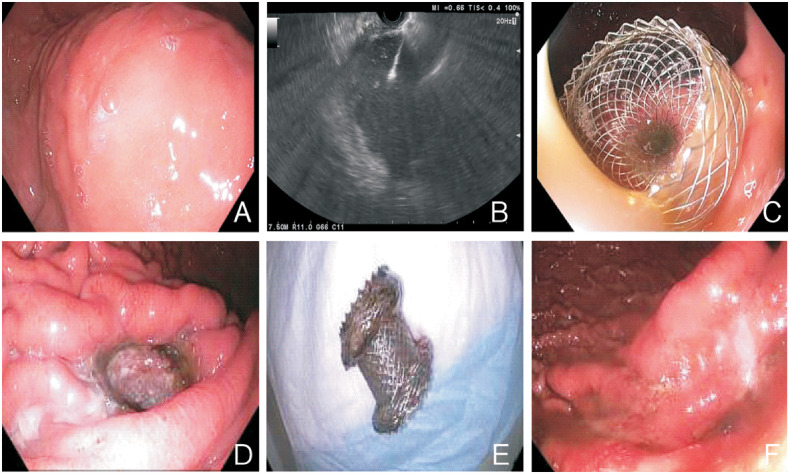
Figure 1.
Illustration of steps in the endoscopic ultrasound-guided drainage of pancreatic-fluid collections. (A) Extrinsic compression by pancreatic cyst. (B) Ultrasound view of cystogastrostomy. (C) Stent at insertion. (D) Stent prior to removal. (E) Retrieved stent. (F) Gastrostomy site after stent removal

