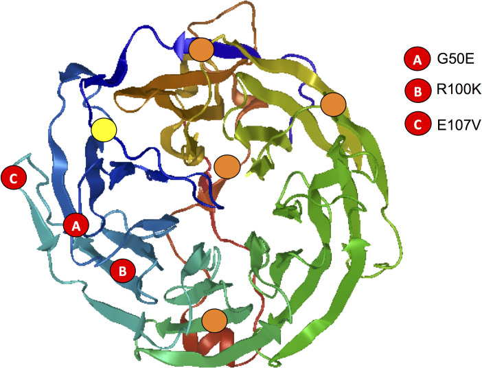
Fig 5. Mutations in PfCoronin conferring resistance to artemisinin are at opposite ends of the putative actin binding sites.
Structure of WD-40 domain of Plasmodium falciparum generated through SWISS-MODEL homology modeling [22] using the crystal structure of the WD-40 domain of Toxoplasma gondii Coronin (4OZU.pdb) [24], which is 43% identical with 99% coverage to the the WD-40 domain of P. falciparum. The predicted actin binding sites are indicated in orange and conserved actin binding site from mouse coronin 1A (R23) [24] indicated in yellow. The mutations that confer artemisinin resistance [17] are indicated in red.

