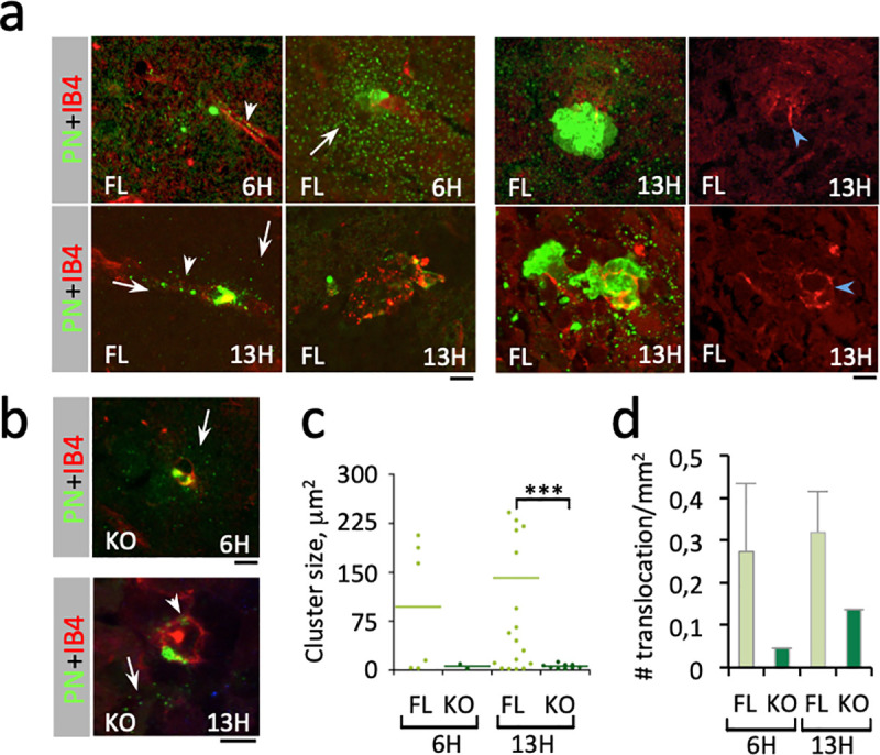
Fig 2. PN translocation across the BBB is increased by aCx43 expression.

Immunofluorescence analysis of brain slices from PN-infected mice. The time post-infection is indicated. Scale bar = 5 μm. a, b, representative micrographs. Red: IB4-labeling of endothelial vessels; green: PN capsule. FL: aCx43FL/FL mice; KO: aCx43-/- mice. Arrows: capsular remnants in the brain cortex. Arrowheads: capsular remnants in vessels. a, right panels: blue arrowheads point at vessel damages. c, size of translocated PN microcolony. 6H: FL, N = 3, 6 foci; KO, N = 3, 2 foci. 13H: FL, N = 3, 20 foci; KO, N = 3, 9 foci). d, frequency of bacterial translocation events per mm2 of brain slice (N = 2, n = 900 60x microscopy fields). c, d, median values are indicated.
