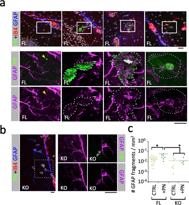
Fig 3. PN induces the aCx43-dependent destruction of astrocytic GFAP network.

Immunofluorescence analysis of brain slices from PN-infected mice. The time post-infection is indicated. Scale bar = 5 μm. a, b, representative micrographs. Red: IB4-labeling of endothelial vessels; green: PN capsule; purple: GFAP. Higher magnifications of the insets in the top panels (a) or left panel (b) are shown. a, FL: aCx43FL/FL mice. Arrow: GFAP association with a single translocated bacterium. b, KO: aCx43-/- mice. c, quantification of GFAP fragments per μm2 in area corresponding to capsular shedding (dotted area) associated with translocated bacterial microcolony. FL CTRL, N = 2, > 10 000 fragments; FL+PN, N = 2, 1658 fragments; KO CTRL, N = 2, 3619 fragments; KO+PN, N = 2, 263 fragments. Mann and Whitney. *: p < 0.05.
