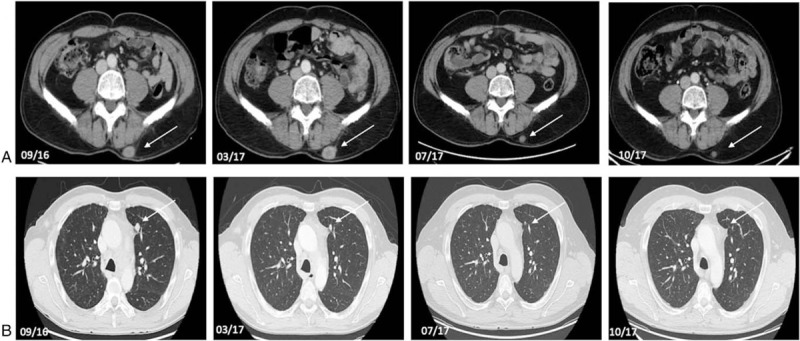
Figure 1.

Axial contrast-enhanced CT-scan of abdomen and chest of a 49-year-old male patient (#1) with a NET G2 of the larynx with pulmonary, osseous, cerebral, cutaneous, and subcutaneous metastases. Axial contrast enhanced CT-scans between September 2016 and October 2017 (09/16, 03/17, 07/17, 10/17) show regression of subcutaneous (a) and pulmonary metastases (b) during therapy with pembrolizumab. Tumor lesions are indicated by white arrows.
