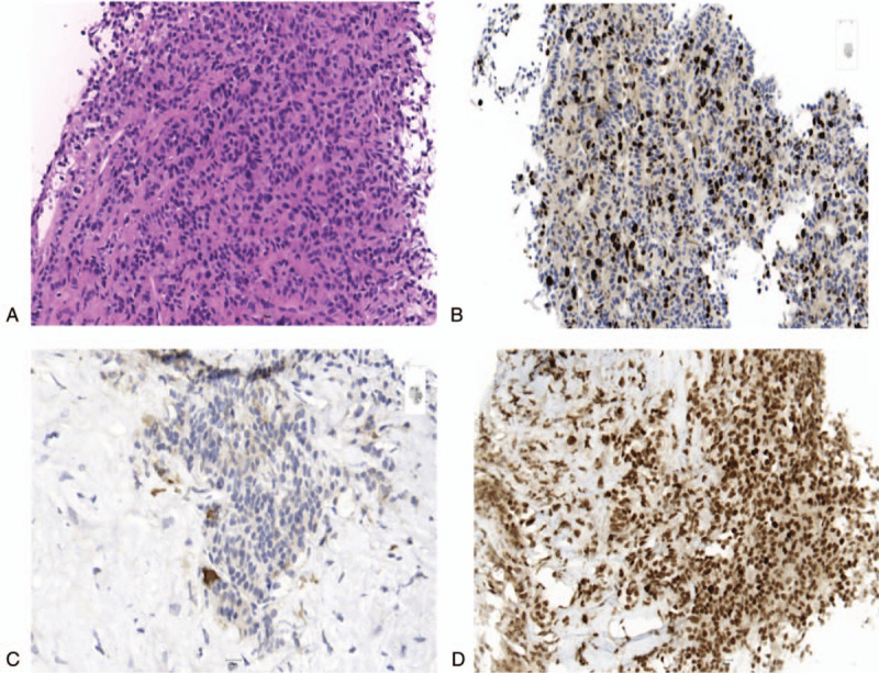
Figure 2.

Immunohistochemical expression of tumor cells of hepatic metastases of a 63-year-old female patient (#2), who was initially diagnosed with a NET G2 of the right kidney, which progressed into a NET G3 during course of disease. H&E stain (20×) of the tumor cells of hepatic metastases shows cells with moderate nuclear pleomorphism, finely speckled chromatin and finely granular eosinophilic cytoplasm (a). Immunohistochemical staining (20×) reveals a Ki-67 proliferation index of 25% (b) few scattered cells with membranous PD-L1 expression (c) and no deficiency in mismatch repair proteins such as MSH6 (d).
