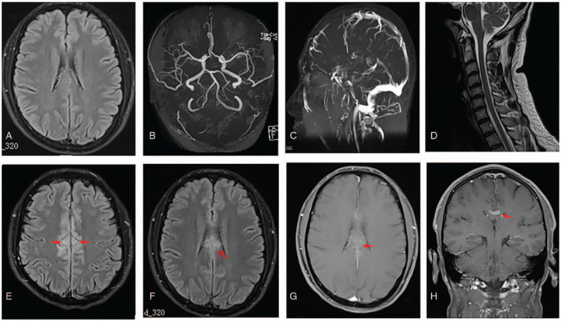
Figure 1.

Magnetic resonance (MR) images of the brain and cervical spine. An initial fluid-attenuated inversion recovery (FLAIR) image (A), brain MR angiography (B), brain MR venography (C), and cervical spine MR images (D). All show no obvious abnormalities. Repeated FLAIR image after the condition worsened, demonstrating an abnormal hyperintense signal in the medial aspect of the bilateral frontal lobes and involvement of the cingulate cortex (E and F, arrows); a gadolinium-enhanced T1-weighted image showing enhancement in the parts above the lesions (G, arrow); and abnormal enhanced meninges (H, arrow).
