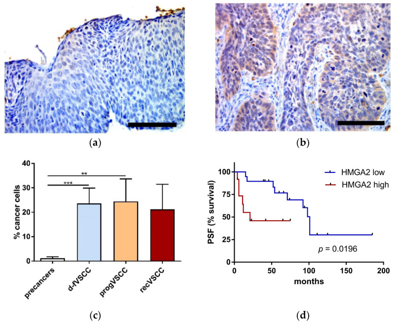
Figure 2.
The percentage of HMGA2-positive neoplastic cells. Examples of immunohistochemical HMGA2 staining performed on tissue sections of high-grade squamous intraepithelial lesions (HSIL) (a) and progVSCC (b) tumor samples. Images were taken at 40× magnification. Scale bar, 100 μm. Comparison of the IHC results for premalignant lesions (HSIL; n = 44 and dVIN; n = 6), d-fVSCC (n = 19), progVSCC (n = 13) and recVSCC (n = 7) samples (c). Survival curve (time to progression) according to the relative numbers of HMGA2-positive cancer cells in primary VSCC tumors (n = 31) (d). Significant alternations are indicated by asterisks (**, p-value ≤ 0.01; ***, p-value ≤ 0.001). Abbreviations: PFS, progression-free survival; high and low, high and low proportion of HMGA2-positive cancer cells (≥30 and ≤10 cells, respectively).

