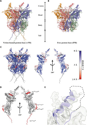
Fig. 4. The HAdV-F41 PB undergoes assembly-induced conformational changes.

(A) Cartoon representation of the virion-bound PB (vPB), which can be divided into a crown, head, body, and tail. (B) Cartoon representation of the free PB (fPB). (C) Cartoon representation of the vPB and a single vPB monomer chain, each colored by Cα RMSD indicating the local degree of difference in Cα positioning between the vPB and fPB structures. (D) Cartoon representation of a single vPB monomer chain (gray). Missing residues in the fPB structure are highlighted in red. (E) Cartoon representation of the helix and disordered loop region containing the integrin-binding IGDD motif (dashed line) located in the crown. The electron density is shown as a transparent surface.
