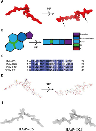
Fig. 5. Location of DNA binding protein V at the interface of the three nonperipentonal hexon subunits.

(A) Surface representation of V electron density. Arrows indicate the positions fixed during bioinformatics analysis. (B) Schematic representation of V and its location in the HAdV-F41 ASU. (C) Alignment of the identified V amino acid sequence from HAdV-C5, HAdV-D26, HAdV-F40, and HAdV-F41. Coloring represents percent sequence identity, with dark blue illustrating 100% homology. (D) Graphical representation of the modeled HAdV-F41 V peptide, shown in maroon, and stick representation covered by the corresponding electron density, shown as transparent surface. (E) Surface representation of the HAdV-C5 [EMD-7034 (20)] and HAdV-D26 [EMD-8471 (21)] electron densities located at the same position in their respective ASUs.
