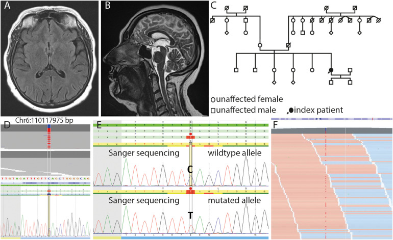
FIGURE 1.
Axial FLAIR (A) and sagittal T2w (B) images show no morphologic features of neurodegeneration or structural damage to the brain. (C) Pedigree of the family. Note that information on clinical symptoms of the patients’ relatives was available only until her age of 20 years. (D) JSI-Sanger data with the heterozygous variant in FIG4 compared to in-house next-generation sequencing (NGS) data, showing this variant in heterozygous state (upper part) and randomized in-house reference pool, which shows the absence of this variant in wildtype (lower part). (E) DNA sequence of the wild-type and mutated allele containing the substitution c.2467 C > T, which leads to the nonsense variant p.Q823* on protein level (red dot) in FIG4. (F) Mapped sequencing reads show the variant on forward (orange) and reverse (blue) strand in approximately 50% of the reads, which is expected for heterozygosity with a sequencing depth (coverage) of more than 500 reads per base.

