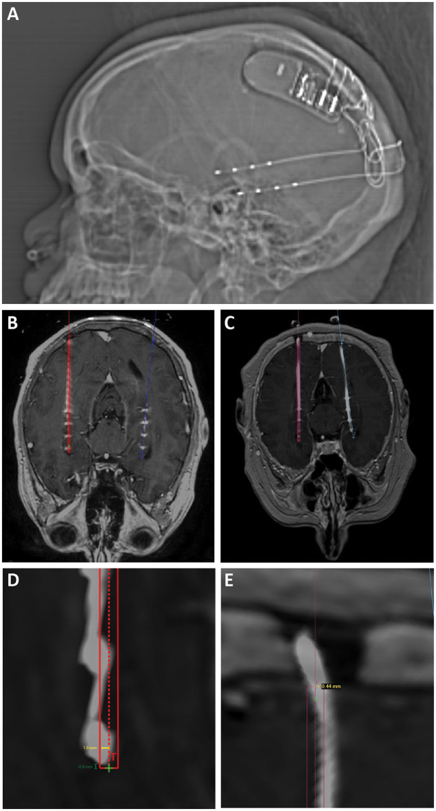
Figure 1.
(A) Scout x-ray showing lateral view after implantation of bilateral hippocampal RNS electrodes and RNS generator. (B) Intra-operative O-arm fluoroscopic CT projected over preoperative planning MRI is used to confirm target accuracy electrodes compared to operative plans (displayed in red and blue). (C) Post-operative CT projected over preoperative planning MRI was also used to confirm target accuracy in cases where O-arm fluoroscopic CT was not performed. (D) Radial target error (yellow line) was measured as the distance from the planned electrode target to the center of the actual electrode position. Depth target error (green) was measured as the difference in depth between the implanted electrode and the planned electrode tip measured along the trajectory of the implanted electrode. Positive values represent electrodes that were implanted past/deeper to target. Negative values represent electrodes that were implanted more shallow compared to target. (E) Radial entry point error was measured as the distance from the planned electrode entry point at the inner table of the skull to the center of the implanted electrode.

