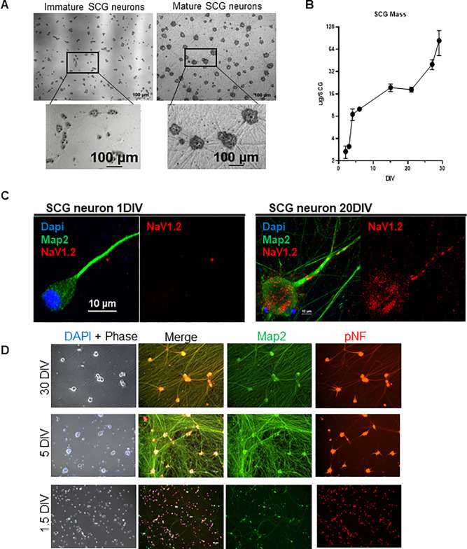
Fig 1. Characteristics of Neuronal Maturation of the Superior Cervical Ganglia.
A: Maturation of SCG neurons is characterized by building a neuronal-network. Phase contrast images comparing immature and mature SCG neurons. Mature neurons develop dense axon bundles (straight lines) and clusters of soma (dark grey). B: Mass per dissociated SCG at various DIV (days in vitro). Bars represent standard deviation. C: Mature neurons express NaV1.2 Immunofluorescence staining in axon. Axon of an immature (1 DIV) SCG neuron does not localize NaV1.2 compared to mature (20 DIV) axon localizing Nav1.2. Map2 serves as a neuronal marker and Dapi for nucleus. D: SCG maturation is acquired by 5 DIV. At 1.5 DIV, the maturation marker pNF (in red) is restricted to the cell body (round) and absent from the axon. At 5 DIV pNF localization spreads to the proximal axon. At 30 DIV pNF is expressed throughout the whole axon as seen by the colocalization of pNF and Map2. Map2 (in green) is a neuronal marker that does not change with age.

