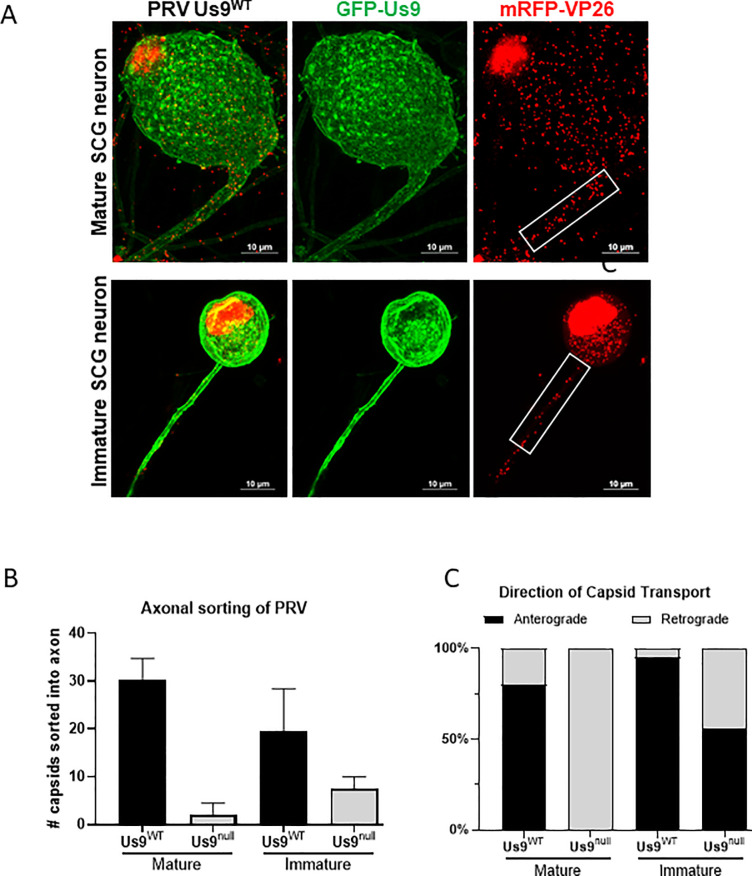
Fig 2. Axonal sorting of Pseudorabies virus depends on neuronal age.
A: Confocal image of SCG neuron infected with PRV expressing mRFP-VP26 (capsid) and GFP-Us9 at 10 MOI for 12 hours. The number of PRV particles, represented by mRFP-VP26 puncta, that sorted into the proximal 30um of axon (white box) are measured. B: Quantification of particles sorted into immature and mature SCG axons. Bars represent standard deviation. C: Live-microscopy quantification measuring the dynamics of particle sorting. Sorted particles were categorized as moving in the anterograde direction (away from cell-body) or retrograde direction (towards cell body).

