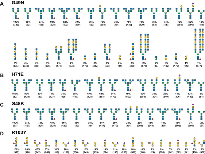
Figure 4.
Oligosaccharide binding specificity of BGL mutant proteins using a mammalian glycan array. Fluorescently labeled proteins G49N, H71E, S48K, and R103Y were used to probe a mammalian glycan array at the Consortium for Functional Glycomics. (A–D) The specificity of each mutant was determined by testing its ability to bind to a printed array (version 5.3) consisting of 600 mammalian glycans. The structures of oligosaccharides bound by each protein are shown in decreasing order of ligand binding efficiency down to 1% binding relative to the strongest signal (see Supplementary Tables 3–6 for raw array data). Structures appearing more than once reflect identical glycans in the array that possess different chemical linkers. The CFG array’s Glycan ID numbers for each structure are shown in parentheses. Sugar symbols are as shown in Fig. 2.

