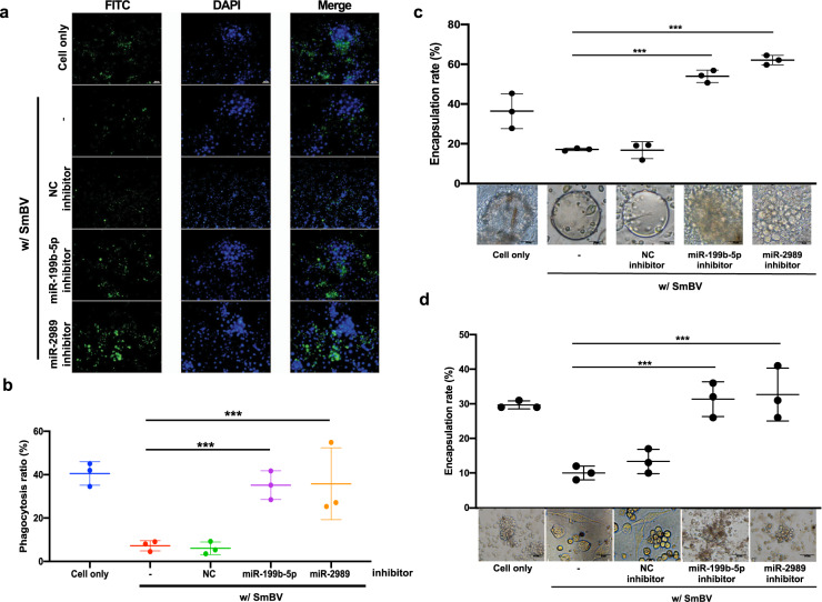
Fig. 3. Inhibition of SmBV miRNA expression increases cellular immune responses in S. litura.
Third-instar S. litura larvae were injected with SmBV and miR-199b-5p or miR-2989 inhibitor; hemocytes were extracted 36 h after the microinjection. a The hemocytes were mixed with FITC-labeled E. coli and the phagocytosis activity of hemocytes was determined by green fluorescence emitted from the ingested E. coli. b The phagocytosis ratio (%) was derived from the proportion of FITC to DAPI; FITC can be seen as green fluorescence detected from FITC-labeled E. coli and DAPI can be seen as blue fluorescence detected from stained hemocytes. (The p-value of miR-199b-5p inhibitor: 0.00548445 and miR-2989 inhibitor: 0.04668200). c Encapsulation assay determining binding of multiple hemocytes to the Sephadex A-25 beads added in the cell culture. The encapsulation rate was calculated by KP assay. Bottom: representative images of one of the Sephadex A-25 beads added in each cell culture. (The p-value of miR-199b-5p inhibitor: 0.00078001 and miR-2989 inhibitor: 0.00019559). d Encapsulation of C.elegans in the hemocoel of S. litura. 24 h after injection, encapsulated nematodes were recovered from S. litura. (The p-value of miR-199b-5p inhibitor: 0.00491668 and miR-2989 inhibitor: 0.00416911) NC inhibitor: negative control inhibitor. All experiments were performed with eight biological replicates (n = 8). Data are expressed as the mean and standard deviation (SD). p-values were calculated using Student’s t-test (*p < 0.05; **p < 0.01; ***p < 0.0005).

