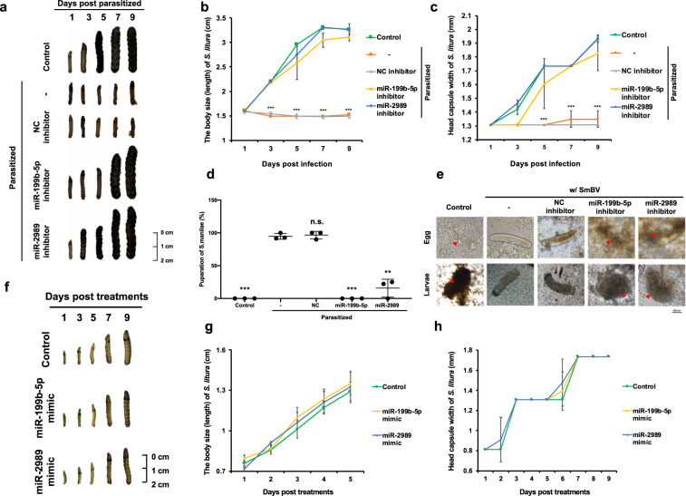
Fig. 5. Inhibition of SmBV miRNA expression decreases S. manilae parasitism rate.
a Third-instar S. litura larvae were microinjected with miRNA inhibitors before parasitism by S. manilae wasps for 48 h. Arrow: S. manilae. The developments of S. litura larvae were measured by their b body sizes (lengths) and c head capsule width after parasitism (Control: unparasitized S. litura) (Green square: Control, orange circle: Parasitized, gray star: Parasitized + NC inhibitor, yellow triangle: Parasitized + miR-199b-5p inhibitor, blue diamond: Parasitized + miR-2989 inhibitor). (The p-value in b on 3–9 days post infection are 0.00000635, 0.00000006, 0.00001210, and 0.00000030) (The p-value in c on 5–9 days post infection are 4.79293E−28, 0.00020260, and 0.00038115). d The pupation rate of S. manilae wasps in S. litura (n = 3) over 10 continuous days. The number of wasps that successfully pupated in the group without miRNA inhibitor injection was set as 100%. (The p-value of Control: 0.00075194, miR-199b-5p inhibitor: 0.00075194, and miR-2989 inhibitor: 0.00561026). e Encapsulation assay determining binding of multiple hemocytes to S. manilae eggs (upper panel) and 4-day-old S. manilae larvae (lower panel) added in the cell culture. Arrow: encapsulated and melanized S. manilae egg or larvae. f Second-instar S. litura larvae were microinjected with miRNA mimics and monitored over a 9 days period post-injection. (Control: S. litura without miRNA mimic injection.) The developments of S. litura larvae were measured by their g body sizes (lengths) and h head capsule width after miRNA mimic injection (Green square: Control, yellow triangle: miR-199b-5p mimic, blue diamond: miR-2989 mimic). All experiments were performed with three biological replicates. Data are expressed as the mean and standard deviation (SD). p-value were calculated using Student’s t-test (***p < 0.005).

