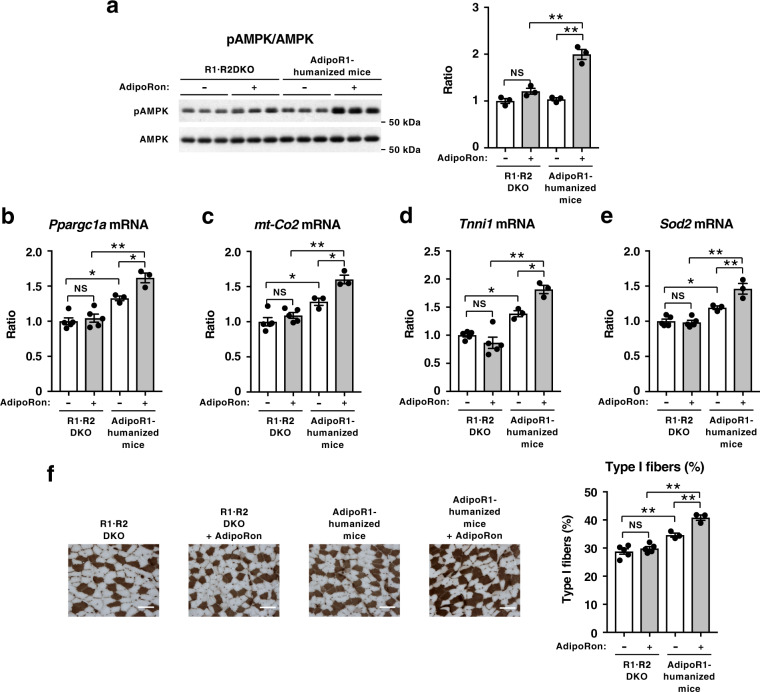
Fig. 5. AdipoRon could increase pAMPK and genes involved in mitochondrial biogenesis as well as oxidative stress-detoxification in AdipoR-humanized mice on a high-fat diet.
Phosphorylation and amount of AMPK (a) in skeletal muscle from R1·R2DKO mice or AdipoR1-humanized mice on a high-fat diet, treated for 7.5 min after injection of 50 mg of AdipoRon per kg body weight intravenously into mice through an inferior vena cava catheter. Ppargc1a (b), mt-Co2 (c), Tnni1 (d) and Sod2 (e) mRNA levels in skeletal muscle from R1·R2DKO mice or AdipoR1-humanized mice on a high-fat diet, treated once daily oral administration of AdipoRon (50 mg per kg body weight) for two weeks. ATPase (pH 4.3 for type I fibers) staining of soleus muscle (f) (scale bars, 100 µM), quantification of type I fibers based on fiber-type analyses in soleus muscle from R1·R2DKO mice and AdipoR1-humanized mice on a high-fat diet with or without AdipoRon (50 mg per kg body weight). All values are presented as means ± s.e.m. *P < 0.05 and **P < 0.01 compared to control mice or as indicated (ANOVA followed by Tukey–Kramer multiple comparison tests). NS, not significant. n = 3 each (a) R1·R2DKO mice (Vehicle: n = 5; AdipoRon: n = 5), AdipoR1-humanized mice (Vehicle, n = 3; AdipoRon, n = 3) (b–f).

