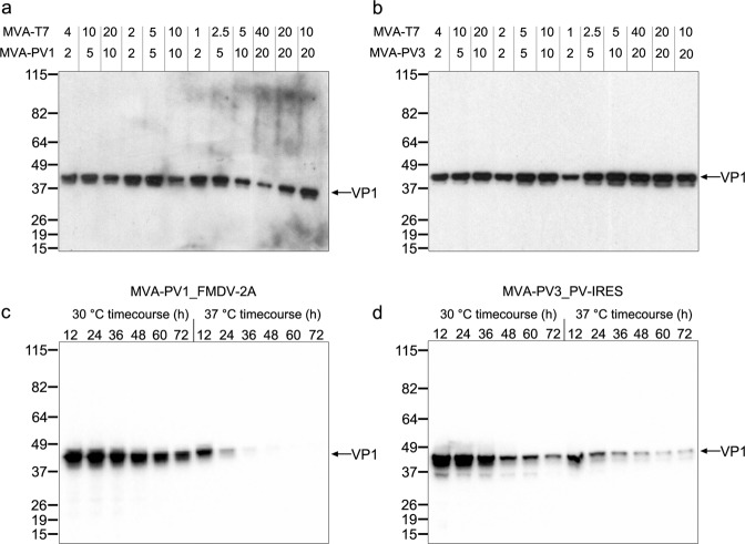
Fig. 2. Small-scale expression of MVA-PV1 and MVA-PV3 constructs to determine optimum parameters for large-scale infection/expression.
a, b Western blots of 6-well screens for MVA-PV1_FMDV-2A (a) and MVA-PV3_PV-IRES (b) co-infected with MVA-T7 to test optimum MOI ratios for expression. MOI ratios tested in each lane are shown above the blot for MVA-T7 and either MVA-PV1_FMDV-2A (MVA-PV1, a) or MVA-PV3_PV-IRES (MVA-PV3, b). c, d Western blots of MVA-PV1_FMDV-2A (c) and MVA-PV3_PV-IRES (d) co-infected with MVA-T7 at their optimum MOIs determined from (a) and (b) to test temperature (30 °C and 37 °C) and time point of harvest (12–72 h, in 12 h time points). Detection was with anti-poliovirus blend of monoclonal antibodies MAB8566 (Millipore).

