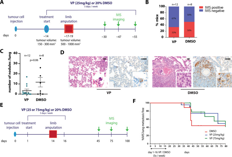
Fig. 6. Verteporfin treatment reduces EwS lung metastasis in a mouse xenograft model.
(a) Experimental setting 1: set-up and VP treatment scheme. Luciferase-expressing TC71 cells were injected into the tibial crest of mice. Intra-peritoneal injections of VP (25 mg/kg) or solvent control (20% DMSO) started once tumours reached a specific size. When tumours reached a volume of 150–300mm3, tumour-bearing limbs were amputated and VP and control treatments (5 days/week) were continued for a maximum of 35 days. (b) Proportions of mice with IVIS-detectable pulmonary metastases per treatment group. (c) Number of histopathologically detectable tumour nodules in lung sections of control- and VP-treated mice based on evaluation of H&E and CD99 stainings. The mean number ±s.e.m. of tumour nodules per condition is shown. P value was calculated by two-tailed Student’s t-test. (d) Exemplary H&E and CD99 stainings showing a reduced size of EwS lung metastatic nodules (200x magnification, inserts: 600x magnification). (e) Experimental setting 2: set-up and VP treatment scheme. As for setting 1 in (A), luciferase-expressing TC71 cells were injected, but VP (25 mg/kg or 75 mg/kg) and control treatments were started one day after tumour cell injection and stopped two days after limb amputation. (f) Lung metastasis free survival of control- and VP-treated mice from experimental setting 2. Although data are not statistically significant, VP-treated mice show a delay in metastatic on-set.

