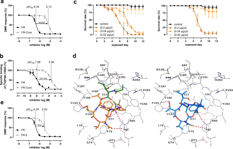
Fig. 4. Evaluation of FR-Core and FR-5 compared to FR.
a Concentration-dependent inhibition of activated Gαq proteins by 1 and 2 as determined by label-free whole-cell DMR biosensing. DMR recordings are representative (mean + s.e.m.) of at least four independent biological replicates conducted in triplicate. b. Competition binding experiments of 1 and 2 versus the FR-derived radiotracer [³H]PSB-15900 at human platelet membrane preparation (50 µg protein per vial), incubated at 37 °C for 1 h. c Exposure of nymphs of a stink bug (Riptortus pedestris) to different concentrations of 1 (left) and 2 (right), survival rate was measured. d Docked poses of 1 (left, represented in sticks and colored in orange, the N-Pp-Hle group present only in 1 is colored in green) and 2 (represented in sticks and colored in blue) in the binding pocket of the Gαq protein shown as line representation. Some of the interactions common for 1 and 2 are indicated by red dotted lines, and the interactions specific for 1 are shown as green dotted lines. Oxygen atoms are colored in red, nitrogen atoms in blue and polar hydrogen atoms in white. e Concentration-dependent inhibition of activated Gαq proteins by 1 and 7 as determined by label-free whole-cell DMR biosensing (see a). Source data are provided as a Source Data file.

