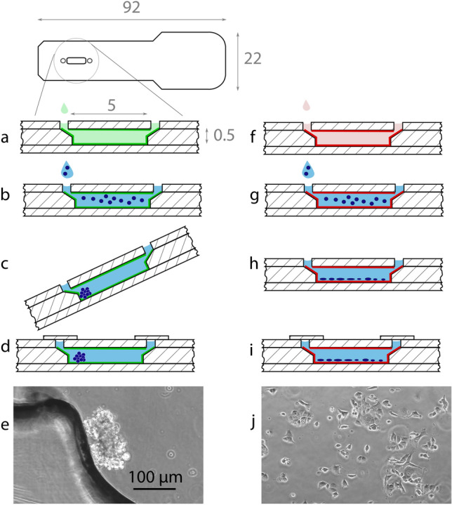
Figure 1.

Schematic of the microfluidic device and sample preparation. The device is coated with pluronic F-127 (a, dimensions in mm) or fibronectin (f). Then, cells are seeded in growth medium (b,g), and incubated for 4 h (c,h). The pluronic-coated device is held at an angle, allowing the cells to settle at the bottom (c). Finally, the devices are sealed with adhesive tape (d,i). Phase-contrast micrograph of the spheroid culture (e) and adherent monolayer (j) of 1800 cells in 2.5 L. (e,j) are the same scale.
