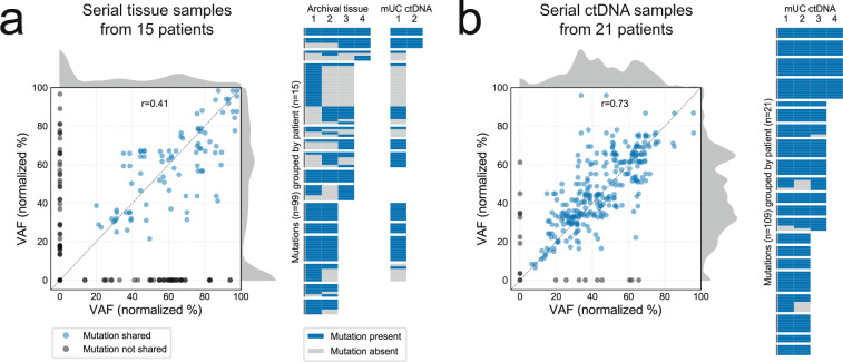
Fig. 3. Temporal heterogeneity in tumor tissue and circulating tumor DNA (ctDNA).
a Mutation detection across same-patient serial tissue samples. Correlation of somatic mutation variant allele fractions (VAFs) in paired tissue samples, with mutations not detected in one member of the pair (VAF = 0) shown in gray (left). Kernel density estimates show a peak in mutations detected exclusively in one sample. Each unique mutation detected in serial tissue is plotted as a row in the heatmap (right), along with their re-detection in ctDNA-positive samples (if available). b Mutation detection across same-patient serial ctDNA samples. Somatic mutation VAFs are strongly correlated (left), with few mutations not consistently detected (right). In both (a) and (b), VAFs are normalized to tumor purity, and those >100 (e.g., on amplified genes) are not shown. Source data are provided as a Source Data file.

