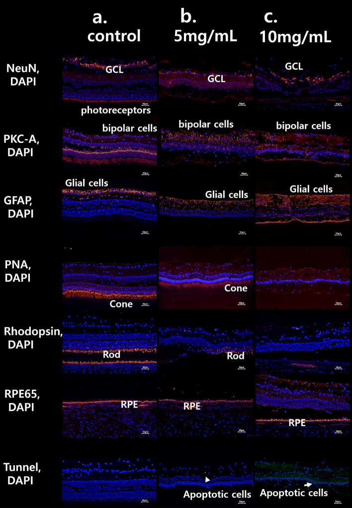
Figure 9.
The immunohistochemistry findings of each representative case of 5 mg/mL MNU group and 10 mg/mL MNU group. (a) The normal immunohistochemistry of a control case. Rhodopsin staining and PNA staining showed well-stained rod and cone cells (each percentage of the stained layer = 15.22% and 14.86%). (b) The immunohistochemistry of a representative case with moderate outer degeneration in the 5 mg/mL group. NeuN staining showed intact ganglion cells and the co-staining of photoreceptors. PKC-α staining showed intact bipolar cells, and GFAP staining showed nearly normal muller cells. Rhodopsin staining showed severely decreased rod cells, and PNA staining showed focal intact cone cells (each percentage of the stained layer = 4.11% and 14.15%). RPE65 staining showed intact RPE and rare co-staining of photoreceptors. There was no apoptotic cell in Tunnel staining. (c) The immunohistochemistry of a representative case with severe outer degeneration in 10 mg/mL group. NeuN and PKC-α stainings showed intact ganglion cells and bipolar cells, respectively. There was no co-staining of photoreceptors in NeuN staining. GFAP staining showed increased staining, especially in the outer retina. Rhodopsin and PNA staining showed relatively rare stained lesions of rod and cone cells (each percentage of the stained layer = 0% and 0%) with co-staining of RPE cells. RPE 65 staining showed intact RPE cells and Tunnel staining showed no stained lesions. RPE retinal pigment epithelium, GCL ganglion cell layer, IPL inner plexiform layer. Each percentage of the stained layers was evaluated by the stained area/all layer area using ImageJ software (1.53a version, National Institutes of Health, Bethesda, MD, USA).

