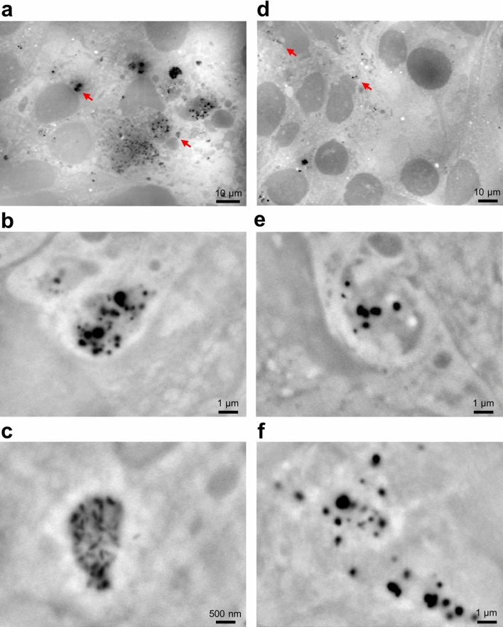
Figure 4.
SE-ADM images of 4T1E/M3 cells 3 and 24 h after the addition of PM2.5. (a) A low magnification (1200×) image of 4T1E/M3 cells 3 h after the addition of PM2.5. A number of PM2.5 aggregates were seen in the whole visual field. (b, c) High magnification (b 10,000×; c 20,000×) images of the areas indicated by red arrows in (a). The images show high-density black particulate aggregates covered with light intracellular membrane-like structures. (d) A low magnification (1000×) image of 4T1E/M3 cells 24 h after the addition of PM2.5. (e, f) High magnification (10,000 ×) images of the areas indicated by red arrows in (d). Scale bars, 10 μm in (a, d), 1 μm in (b, e, f), and 500 nm in (c).

