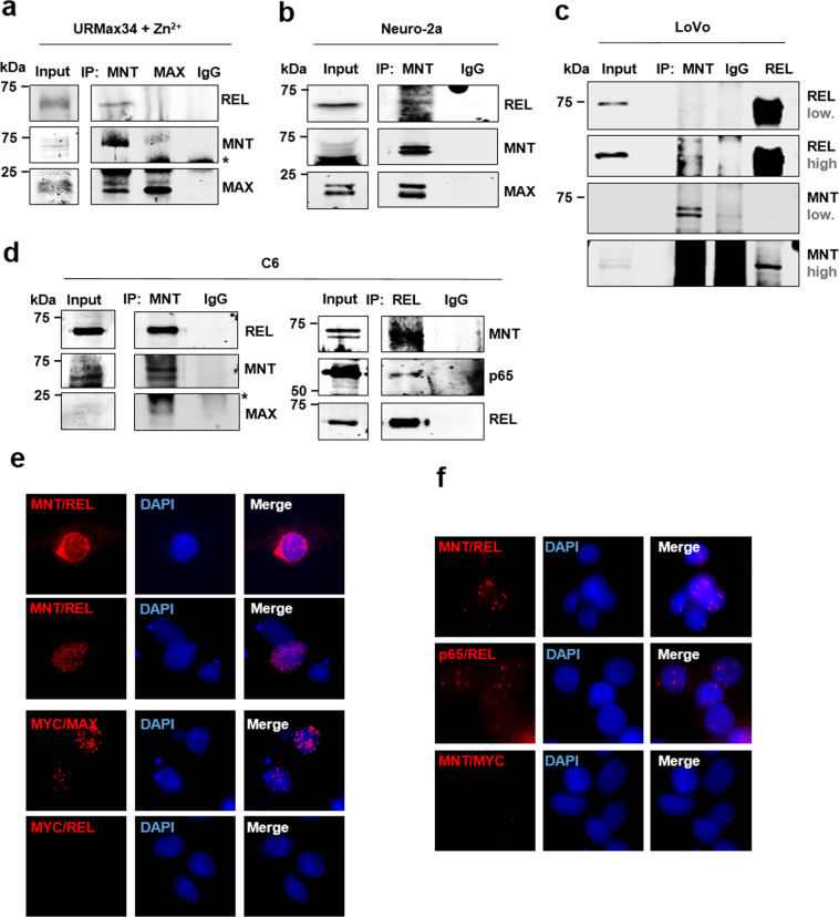
Fig. 2. MNT and REL physically interact.
a Co-immunoprecipitation assay with anti-MNT and anti-MAX antibodies in URMax34 cells after Zn2+ treatment. MAX was used as a positive control for MNT immunoprecipitations. The asterisk marks the position of the heavy IgG band. b Co-immunoprecipitation assay with anti-MNT in mouse Neuro-2a cells. c Co-immunoprecipitation assay in human LoVo cells with anti-MNT and anti-REL. Two immunoblot images of the blots with high and low intensity are shown. Note that the hypotonic buffer required for MNT–REL co-immunoprecipitation does not extract efficiently MNT protein. d Co-immunoprecipitation assay in rat glioma C6 cells with anti-MNT and anti-REL antibodies. The asterisk marks the position of the light IgG band. e rpoximity ligation assay (PLA) performed in C6 cells 48 h after transfection with WT MNT-HA and REL-flag overexpressing vectors. Antibodies anti-MNT/anti-REL, anti-MYC/anti-MAX (positive control), and anti-MYC/anti-REL (negative control). The PLA-positive signal in red and DAPI as a nuclear marker. f PLA in LoVo cells (untransfected cells) with anti-MNT/anti-REL, anti-p65/anti-REL (positive control), and anti-MNT/anti-MYC (negative control) antibodies. The PLA-positive signal is in red and DAPI staining was used as a nuclear marker.

