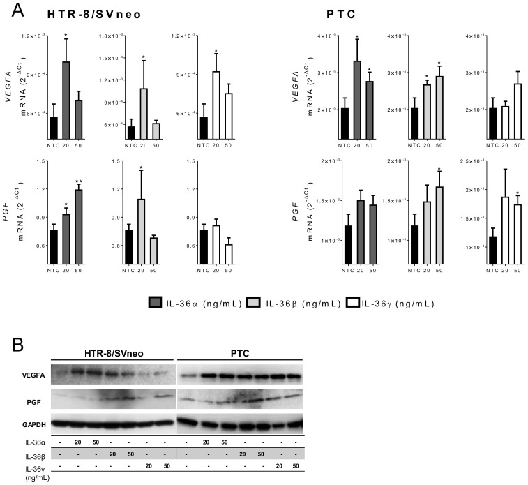
Figure 5.
IL-36 (α, β, and γ) induced expression of angiogenic factors. (A) HTR-8/SVneo and primary trophoblast cells (PTC) were stimulated with 20 or 50 ng/mL IL-36 (α, β, and γ) for 24 h. The mRNA levels of VEGFA and PGF were determined by quantitative real-time PCR and normalized to GAPDH using the 2−ΔCt method. Results from three independent experiments are shown as mean ± SEM. NTC: Non-treated cells. Unpaired two-tailed Student’s t-test with Welch´s correction. * p < 0.05, ** p < 0.01 for significant differences to NTC controls. (B) VEGFA, PGF, and GAPDH proteins were detected by Western blotting using 20 μg cell lysate from HTR-8/SVneo and PTC stimulated for 24 h.

