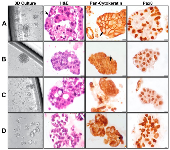
Figure 3.
Morphology of platinum-resistant organoids in culture and their respective histology. (A–C) Organoids from three independent patients with platinum resistant high-grade serous carcinomas. (D) Organoids derived from platinum resistant ovarian cancer cell line OVCAR8. All four organoid models were established in Matrigel-based adherent 3D cell culture. Hematoxylin and eosin (H&E) staining show clusters of malignant cells with pleomorphic nuclei, mitoses (arrow), and necrosis (in A panel). Pan-cytokeratin staining show positivity in the narrow rims of the cytoplasm surrounding the pale staining nuclei (arrow), confirming epithelial origin of the cells. Pax8 shows nuclear staining of the malignant cells, consistent with Mullerian origin of the tumors. The morphological features and the immunohistochemical reactions are remarkably similar in all organoid cultures. All 3D culture organoid pictures are taken with a 20 × objective. All microscopic pictures are taken with a 60 × objective.

