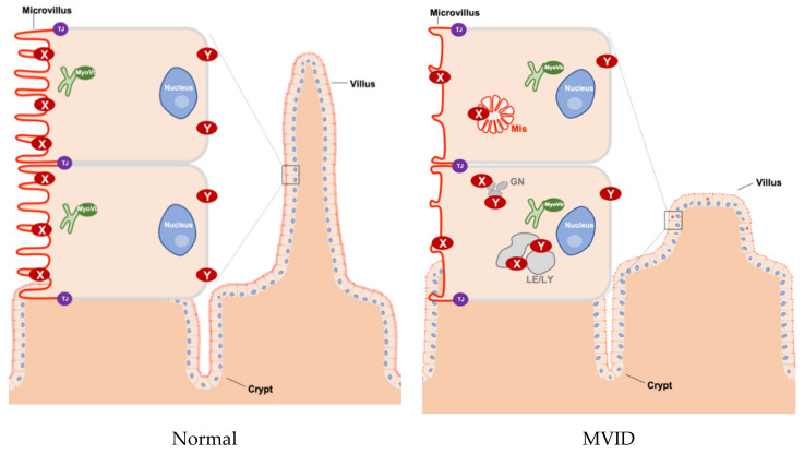
Figure 1.
Schematic overview of tissue and cellular characteristics of healthy and MVID intestinal epithelium. In healthy enterocytes, the recycling endosome (green) is located sub-apically and plays an important role in transporting proteins to the plasma membrane (in particular to the apical membrane). However, in MVID that is caused by the loss function of myosin Vb (MyoVb), the villi show hypoplasia and microvilli are atrophic. Moreover, the proteins (marked as X and Y) in the plasma membrane are mislocalized in microvillus inclusions (MIs) or in enlarged late endo/lysosomes (LE/LY) and granules (GN). X indicates CD10, DRA, GLUT5, NHE3, SGLT1, sucrase-isomaltase (SI), 5’-nucleotidase (5′NT), alkaline phosphatase (AP), AQP7, CD36, CFTR, DPPIV. Y indicates transferrin receptor (Tfr), Na+/K+ ATPase. TJ: tight junction, MIs: microvillus inclusions, LE: late endosomes, LY: lysosomes, GN: granules.

