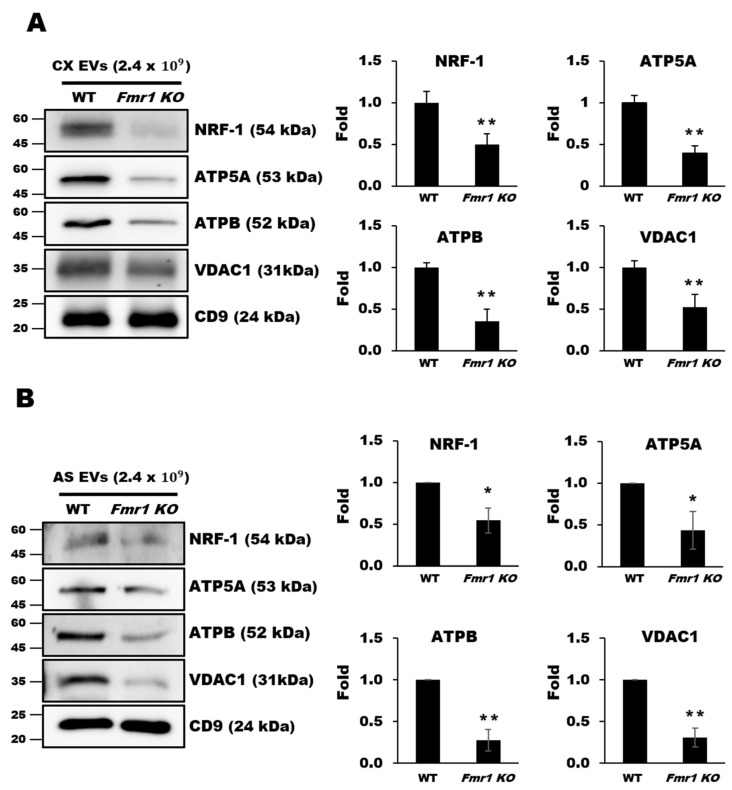
Figure 6.
Depletion of mitochondrial components in extracellular vesicles (EVs) from cerebral cortices (A) and astrocytes (B) of Fmr1 KO mice. The expression of mitochondrial proteins including NRF1, ATP5A, ATPB, and VDAC1 was analyzed by western blot analysis of EVs isolated from the cortices (CX) (A) and astrocytes (AS) (B) of WT and Fmr1 KO mice. Fold differences in expression were determined by measuring band densities with NIH ImageJ software. CD9 was used as an EVs marker and loading control. Each value represents the mean ± SD of three independent experiments. * p < 0.05; ** p < 0.01 vs. WT mice.

