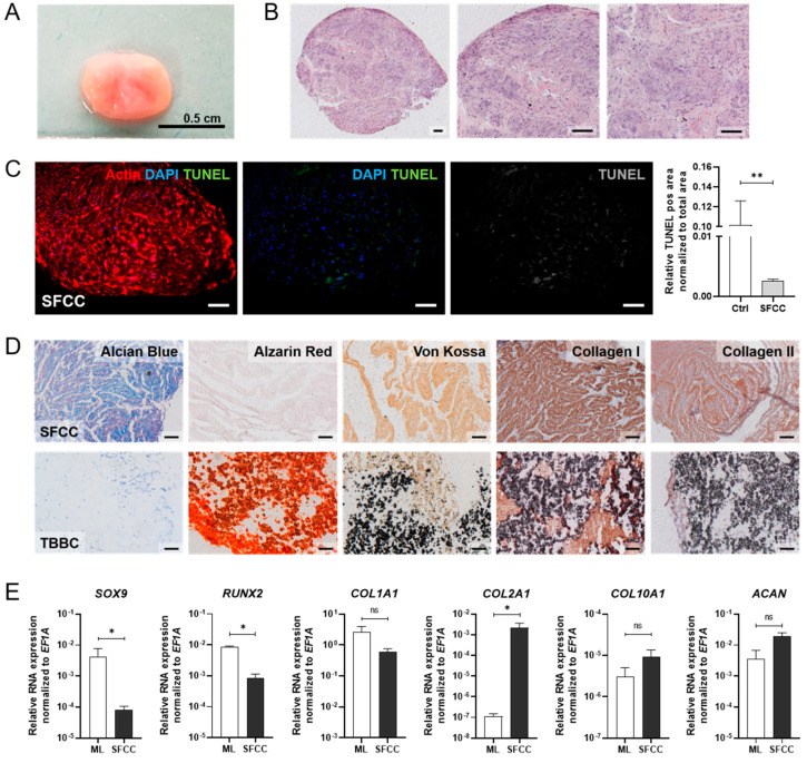
Figure 5.
In vitro studies on the 3D scaffold-free cartilage constructs (SFCC) based on human MSCs. (A) Macroscopic overview of the in vitro 3D SFCCs. (B) Histological evaluation of the morphology via H&E staining. Exemplary images for n = 6. Scale bars indicate 200 µm. (C) Detecting apoptotic cells (green) using TUNEL staining after 21 days without mechanical force. Exemplary image for n = 4. Scale bars indicate 200 µm. Pos. ctrl = incubation with DNase I for 10 min. (D) Histological (Alcian Blue, Alizarin Red, von Kossa) and immunohistochemistry staining (collagen type 1 and collagen type 2) of the SFCC in comparison with the tricalcium phosphate-based bone component (TBBC) control. Exemplary images for n = 4. Scale bars indicate 200 µm. (E) Total RNA extraction was performed from 3D cultures after 21 days. Gene expression was normalized to the housekeeper gene EF1A. Data are shown as mean ± SEM (duplicates per gene) for n = 3–6. Mann-Whitney U-test was used to determine the statistical significance; p-values are indicated in the graphs with * p < 0.05, ** p < 0.01 (ns = not significant). MSC, mesenchymal stromal cell; EF1A, eukaryotic translation elongation factor 1 alpha.

