Figure 3.
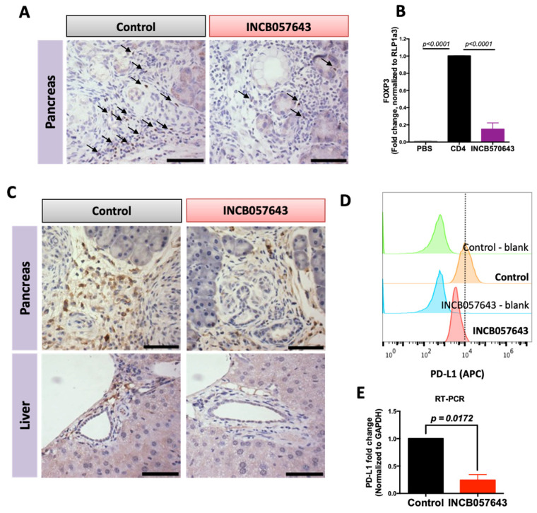
INCB057643 reduces the expression of FOXP3 in CD4 T cells and PD-L1 expression in macrophages. Immunohistochemistry for FOXP3 (A) or PD-L1 (C) in pancreas and liver of KPC mice treated with vehicle control or INCB057643 for 16 weeks. Scale bar = 30 µm. (B) CD4 T cells were isolated from a spleen of a wild type mouse using negative magnetic bead selection. CD4 T cells were plated with anti-CD3 (or PBS, a control for non-stimulated T cells), anti-CD28, IL-2 and TGF-β for 24 h prior to adding INCB057643 (100 nM) for 4 days. CD4 T cells were collected and levels of FOXP3 were determined by PCR. p < 0.001 vs. control CD4 cells in 2 independent experiments. (D) RAW 264.7 macrophage-like cells were treated with conditioned media from PanAsc 2159 cells; representative histograms for levels of PD-L1 analyzed by flow cytometry after treatment with 0.5 μM INCB057643 for 24 h are shown (n = 2) and confirmed by RT-PCR (n = 3) (E).

