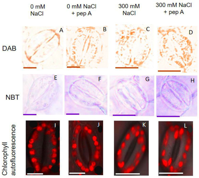
Figure 9.
Hydrogen peroxide and superoxide radical generation and chlorophyll autofluorescence in GCs of quinoa in response to salt stress and pepstatin A. (A–D): 3,3′-Diaminobenzidine (DAB) staining to detect H2O2 in GCs. (E–H) Nitro blue tetrazolium (NBT) staining to detect superoxide radicals in GCs. Chlorophyll autofluorescence in GCs (I–L) was visualized using a laser scanning confocal microscope (LEICA SMD FLCS) at an excitation wavelength of 488 nm and chlorophyll autofluorescence was detected between 629 nm and 697 nm. Bar = 10 µm.

