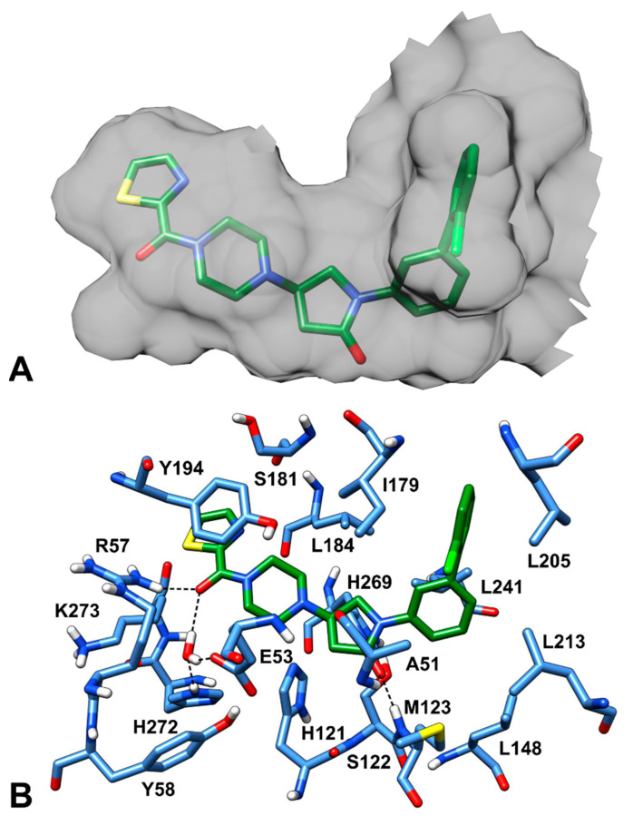
Figure 1.
Piperazinyl-pyrrolidine inhibitor 3l in the MAGL binding site (PDB code: 5ZUN). (A) The inner surface of the protein surrounding the ligand (green sticks), displaying the shape of the ligand-binding cavity, is shown in gray. (B) The protein residues surrounding the ligand, constituting the binding site, are show in blue sticks, while hydrogen bonds are shown as black dashed lines.

