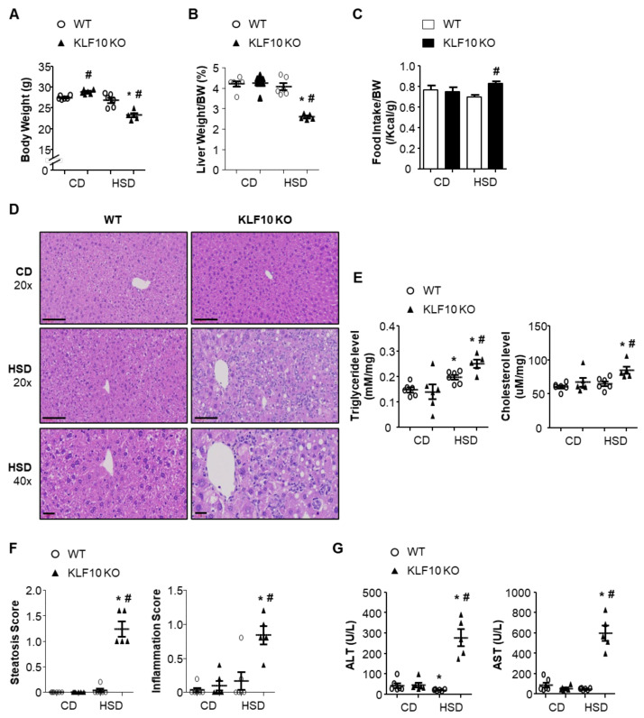
Figure 1.
Klf10 KO mice develop HSD-induced liver injury. Eight-week-old WT and Klf10 KO mice were fed either CD or HSD for eight weeks (n = 5–6 mice/group). (A) Body weights and (B) liver weights were determined after eight weeks of either diet. (C) Food intake (average over seven days) was measured in WT and Klf10 KO mice. (D) Representative histological images of H&E-stained liver sections (Scale bars: 60 μm, Magnification: 20×; enlarged images in the bottom panels of D, 40×). (E) Triglycerides and cholesterol in the liver were measured from liver samples. (F) The grade of the steatosis and inflammation score was calculated. (G) Plasma ALT and AST levels were measured as liver injury markers. All data are representative of at least three independent experiments and expressed as mean ± SEM. Charts were produced using GraphPad Prism 5.0. Statistical differences were determined by two-way ANOVA with Mann–Whitney U test using SPSS v17.0. * p < 0.05 vs. genotype-matched, CD-fed group and # p < 0.05 vs. diet-matched, genotype control. WT, wild type; KO, knockout; CD, control chow diet; HSD, high-sucrose diet.

