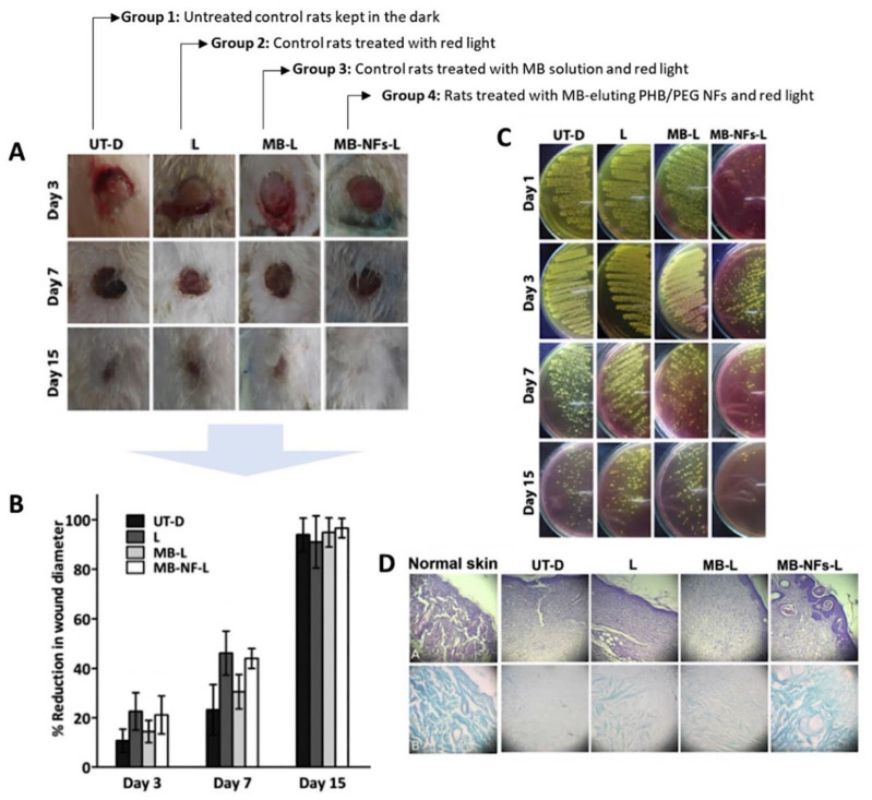
Figure 7.
Healing evolution of different skin wound treatment groups. (A) Morphological characteristics of excision-infected wounds in different animal groups; (B) Time-course of percentage reduction in wound diameter; (C) Degree of wound bacterial contamination over 15 days; (D) Histopathological examination of H & E (top row) and Masson’s trichrome (bottom row) stained excision wounds after 15 days of wounding (figure adapted from [12] with permission of Elsevier).

