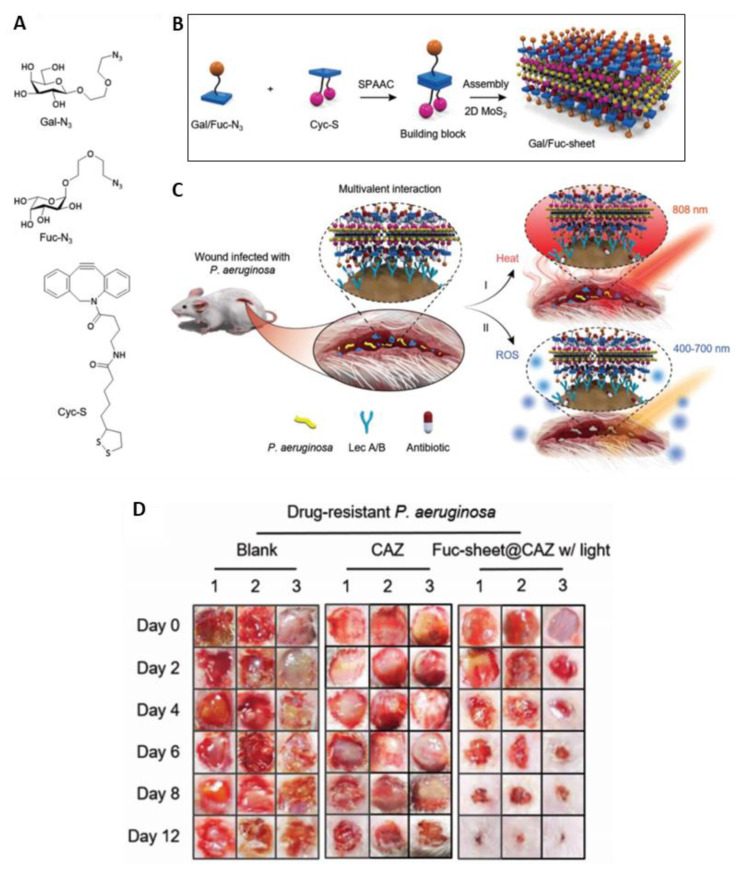
Figure 32.
(A) Structure of azido-galactoside (Gal-N3), azido-fucoside (Fuc-N3), and α-lipoic acid-coupled cyclooctyne (Cyc-S); (B) Schematic illustration of glycosheet formation; (C) Double light-driven therapy of wound infected by P. aeruginosa; (D) Photographs of wounds treated in the presence of glycosheets for multidrug-resistant P. aeruginosa (figure adapted from [88] with permission of John Wiley and Sons).

