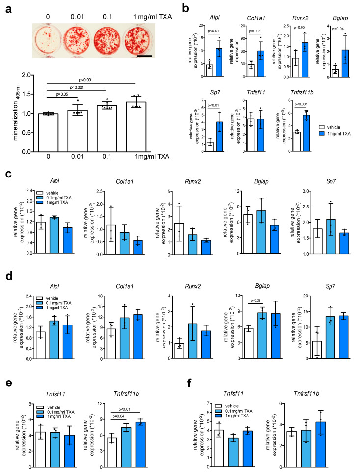
Figure 2.
TXA promotes extracellular matrix mineralization. (a) Representative alizarin red stainings of bone marrow-derived osteoblasts from WT mice differentiated in the presence of indicated concentrations of TXA for 10 days in osteogenic medium. Scale bar 10 mm. The quantification of extracellular matrix mineralization is indicated below. (b) qRT-PCR expression analysis for the indicated genes in bone marrow-derived osteoblasts at day 10 of osteogenic differentiation, stimulated with TXA (1 mg/mL) during the entire course of cell differentiation. (c,d) qRT-PCR expression analysis for the indicated genes in bone marrow-derived osteoblasts at day 2 (c) and day 10 (d) of osteogenic differentiation, stimulated with TXA for 6 h at the indicated concentrations after serum starvation overnight. (e) qRT-PCR expression analysis for the indicated genes in the same cells at day 2 or (f) day 10 of differentiation. For (a–f), n = 3–4 independent cultures per group were used. Data presented are means ± SD. Gene abbreviations: runt-related transcription factor 2 (Runx2), osterix (Sp7), alkaline phosphatase (Alpl), alpha-1 type I collagen (Col1a1), osteocalcin (Bglap), sclerostin (Sost), Rankl (Tnfsf11), osteoprotegerin (Tnfrsf11b).

