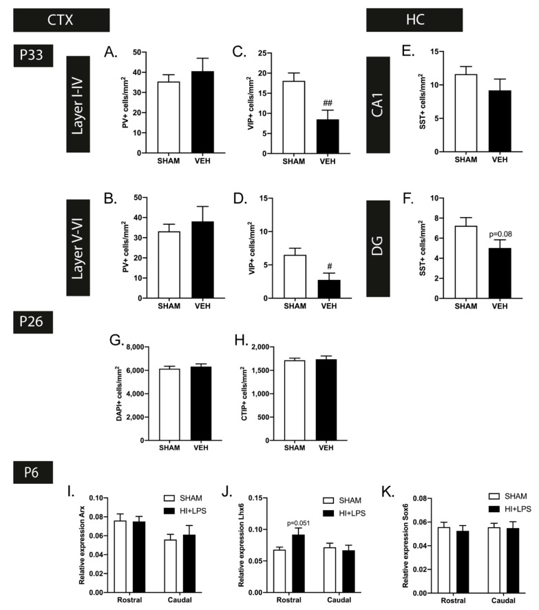
Figure 5.
A selection of developmental disturbances in interneurons persist with age. (A,B) At P33, the observed changes in PV+ cell density were restored in both the upper (A) and lower (B) cortex (SHAM n = 12, VEH n = 9). (C,D) In contrast to the excess in VIP+ cortical interneurons at P26, a decrease in VIP+ cells per mm2 was observed in the upper (K) and lower (L) layers of the cortex of HI+LPS mice at P33 (SHAM n = 12, VEH n = 8). (E,F) The observed SST+ interneuron deficiency in the dentate gyrus (F) of HI+LPS mice showed persistence up to P33 (statistical trend), while the hippocampal CA1 region (E) remained unaffected (SHAM n = 11, VEH n = 8). (G,H) In contrast to the FIPH rat model, cortical cell density, measured by DAPI+ cells/mm2 (G) and CTIP+ cells/mm2 (H), was not affected at P26 by HI+LPS (SHAM n = 12, VEH n = 12). (I–K) Expression of Lhx6 (J) was increased (statistical trend) in the rostral cerebrum of EoP (HI+LPS) animals (n = 4) compared to sham-controls (n = 5) at 24 h after HI+LPS. Induction of EoP did not affect expression of Arx (I) and Sox6 (K). #: p < 0.05; ##: p < 0.01 HI+LPS animals vs. sham-controls; nearly significant p values are indicated in (F,J).

