Figure 3.
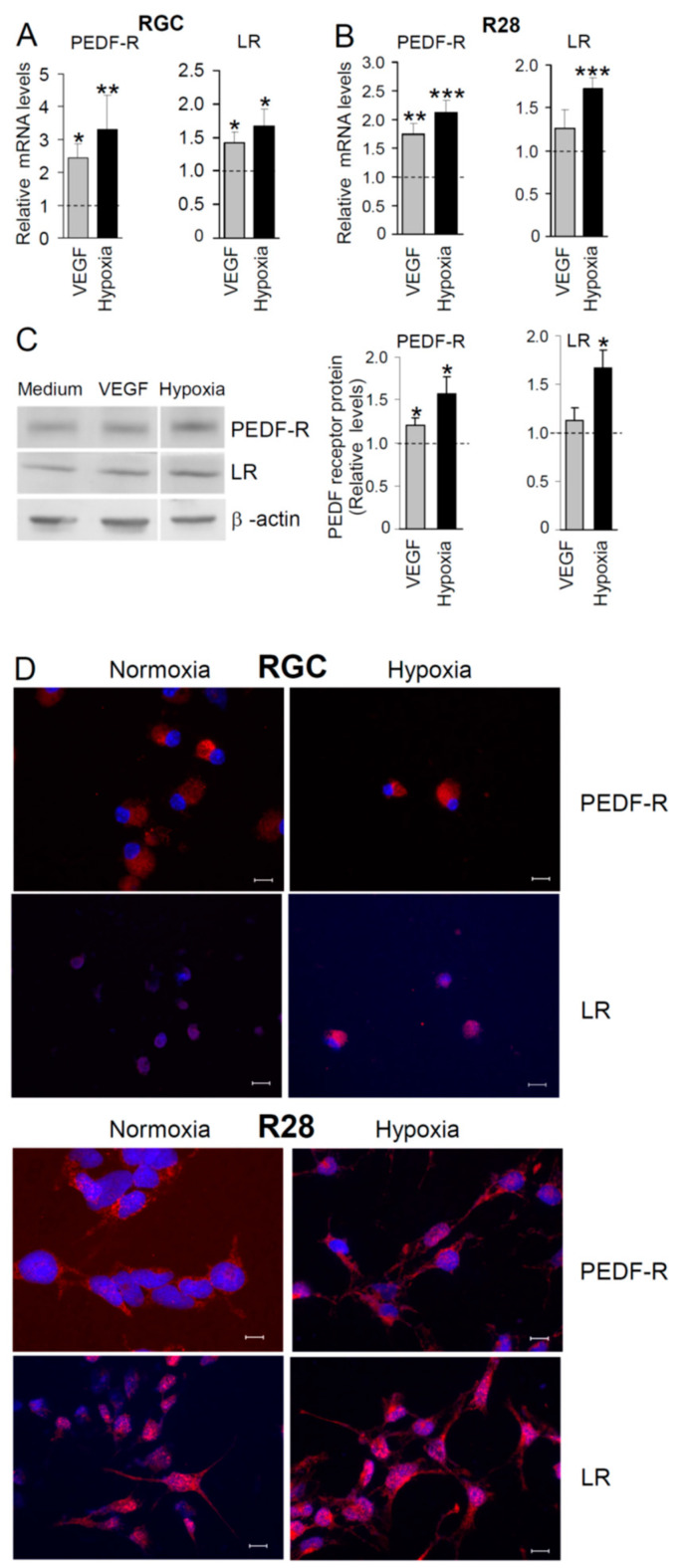
PEDF-R and LR mRNA expression in retinal neuronal cells is regulated by VEGF and hypoxia. Cells were treated with 50 ng/mL VEGF or incubated at 0.2% O2. At 24 h post-treatment, total RNA was prepared from (A) RGC or (B) R28 cells, and expression of PEDF receptors was analyzed by semi-quantitative real-time PCR. Shown are relative mRNA levels of PEDF-R (A, n = 8; B, n = 6–9) and LR (A, n = 6–9; B, n = 5–9). (C) Regulation of PEDF-R and LR proteins in R28 cells is demonstrated by a representative Western blot (left panel). Graphs summarizing data from independent experiments (n = 4–6) are shown (right panel). Results are expressed as relative values (fold change in PEDF-R or LR expression; * p < 0.05, ** p < 0.01; *** p < 0.001; means ± SEM) compared with cells of unstimulated (normoxic) control cultures (dashed lines). (D) RGC and R28 cells cultured under normoxia or hypoxia were stained with antibodies directed to PEDF-R and LR (red fluorescence) and immunofluorescence was detected using a fluorescent microscope. There was no staining in the control samples stained with non-immune mouse IgG1 or rabbit IgG instead of primary antibodies. Cell nuclei were labeled with DAPI (blue fluorescence). Scale bars, 10 µm.

