Figure 2.
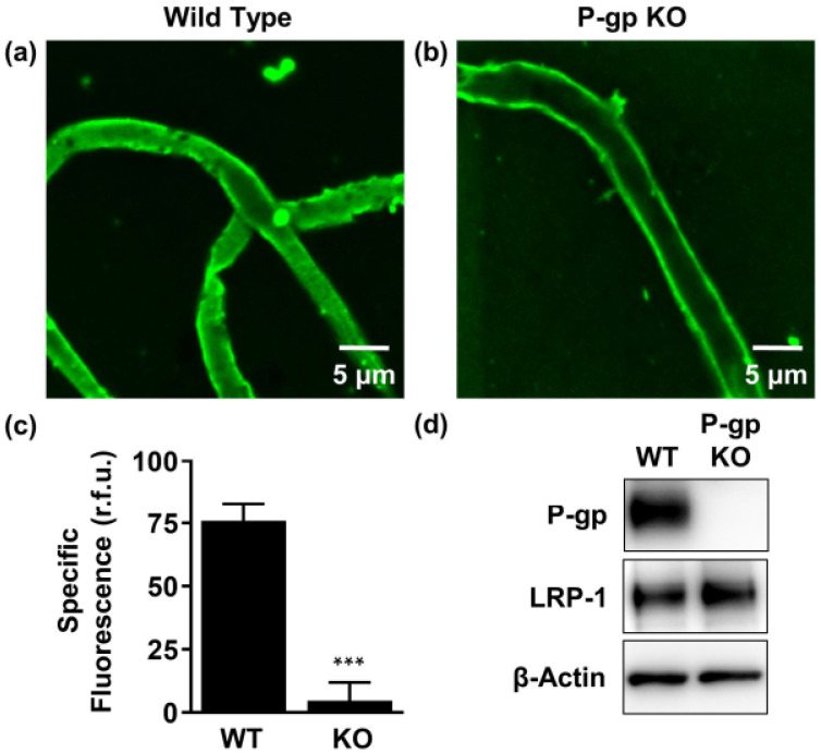
P-gp-mediated human Aβ42 (hAβ42) transport in isolated brain capillaries. (a), (b) Representative confocal images showing accumulation of HiLyteTM-hAβ42 in capillary lumens isolated from wild-type (WT) mice, but not in capillaries from P-gp knockout (KO) mice after a 1-h incubation (steady state; 5 μM HiLyteTM-hAβ42). (c) Data after digital image analysis using ImageJ. Specific fluorescence refers to the difference between total luminal HiLyteTM-hAβ42 fluorescence and HiLyteTM-hAβ42 fluorescence in the presence of the P-gp-specific inhibitor PSC833 (5 μM). (d) Western blot showing P-gp protein expression in isolated capillaries from WT mice, but not in capillaries isolated from P-gp KO mice. In contrast, LRP-1 is expressed in isolated capillaries from both WT mice and P-gp KO mice. β-actin was used as the loading control. Statistics: data per group are given as mean ± SEM for 10 capillaries from one preparation (pooled tissue: WT (n = 10 mice), P-gp KO (n = 10 mice)). Shown are relative fluorescence units ((r.f.u.) scale 0–255). *** Significantly lower than control, p < 0.001.

